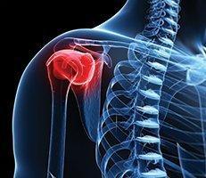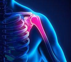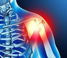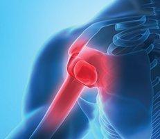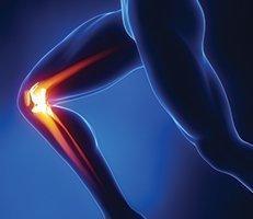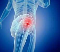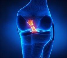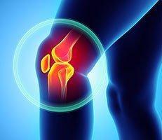
Normal knee function requires a smooth gliding articular cartilage surface on the ends of the bones. The cartilage acts to distribute force during repetitive pound-like movements, such as jumping or running on hard surfaces. Despite its strength, cartilage is vulnerable to damage that can cause chronic knee pain. Injuries take the form of lesions, holes, or divots in the surface of the cartilage.
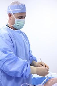
Beverly Hills orthopedic surgeon, Dr. Eric Millstein, specializes in cartilage injuries and treatment. He uses the most advanced cartilage repair surgical techniques to provide his patients with the best results possible.
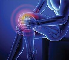
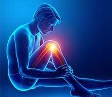
Knee Cartilage Injury Symptoms
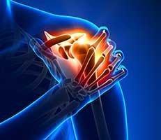
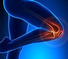
A severe knee cartilage injury can radically change an active adult’s lifestyle. Symptoms of an injury can include locking or catching of the knee as well as localized pain and swelling. This often affects an individual’s ability to work, play, and perform everyday activities. If left untreated, it can hinder your ability to move free from pain, and cause deterioration to the joint surface.
Autologous Chondrocyte Implantation (ACI)
Although damaged cartilage cannot repair or restore itself, advanced surgical technology allows cartilage cells to be harvested from your knee and cultured and multiplied. Restoring articular cartilage can not only relieve pain, but allow better function as well. Most importantly, it can delay or prevent the onset of arthritis.
The biological repair of the cartilage is known as autologous chondrocyte implantation, or ACI. It is a two-step procedure where new cartilage cells are grown and then implanted in the cartilage defect. The procedure restores articular surface and regenerates hyaline cartilage, without compromising the integrity of healthy tissue or the subchondral bone. When you successfully complete ACI and rehabilitation, you should be able to resume all normal activities.
ACI is most useful for younger patients who have single defects larger than 2cm in diameter. There is no danger of a patient rejecting the tissue, as the patient´s own cells are being used.
OATS Repair
Osteochondral Autograft Resurfacing (OATS) is a therapy that utilizes the transplantation of an individual’s own healthy hyaline cartilage from one area to another. The treatment is primarily performed upon traumatic and chronic injury, and is performed using small instruments through incisions on the sides of the knee. A plug of cells are moved from a non-load bearing area of bone and implanted into the damaged joint location. In order to see inside the joint and guide the instruments, Dr. Millstein uses a small video camera called an arthroscope. Due to a limit in the number of harvest sites available, the procedure is best for small defects that are less than 15-20mm in size.
Osteochondral Allograft Transplantation
If a cartilage defect is too large for an autograft, an allograft is usually considered. An allograft is a block of cartilage and bone that can be shaped to fit the exact contour of the defect and then fit into place.
Allografts are typically done through an open incision, and available instruments can create grafts between 10 and 35mm diameter. First, the defect is exposed and sized. A guide pin is then placed through the center of the lesion, vertical to the joint surface. Diseased cartilage and small amounts of bone are removed, and depth measurements are taken from the prepared recipient site. The allograft plug is then removed from the donor tissue, and recipient depth measurements are marked on the plug. Once this is done, excess bone is removed, which in turn creates an osteochondral graft matching the size and depth of the recipient site. Blood and debris are removed, and bony edges are trimmed to facilitate insertion. Lastly, the graft is gently inserted, and loose grafts are fixed with absorbable pins or screws.
Although there are several reparative and restorative options for cartilage replacement, allografting remains the only biomimetic technique that restores architecturally appropriate, mature hyaline cartilage.
DeNovo Graft
DeNovo grafts are tissue grafts used in cartilage repair, and consist of cartilage tissue collected from donors. However, sometimes, the cartilage tissue is grown in a laboratory using human donated cartilage cells. DeNovo is most commonly used for contained, isolated defects on the patella, femoral trochlea, or medical femoral condyle.
DeNovo ET is an engineered tissue graft cultured in a laboratory, using cells collected from juvenile donors. Juvenile cartilage cells are used because they have a greater potential of regeneration than the adult cartilage cells. The graft can be placed over the defect area using a simple single-step surgical procedure.
DeNovo NT, on the other hand, contains natural articular cartilage living cells, harvested from juvenile donors. Transplantation of DeNovo NT is a simple single-step procedure where the graft is fixed on the defect area using fibrin glue.If you have damaged cartilage, schedule a consultation with Dr. Millstein today to find out what cartilage repair treatment will be the best for your condition.
If you have damaged cartilage, schedule a consultation with Dr. Millstein today to find out what cartilage repair treatment will be the best for your condition.
Beverly Hills-based board certified orthopedic surgeon Dr. Millstein is highly experienced and trained in using the most advanced surgical techniques available today. If you want to make sure that ACI is right for you, please schedule a consultation with Dr. Millstein by calling (310) 595-1030.